BUDA-gSlider for high-resolution, distortion-free diffusion imaging
We are pleased to announce the release of our BUDA-gSlider sequence and off-line reconstruction packages for high-resolution, distortion-free diffusion imaging. This package is developed by research groups at Martinos Center for Biomedical Imaging / MGH and Stanford University.
Pulse sequence for BUDA-gSlider is currently available for the Siemens Scanners on the VE11C platform. The off-line reconstruction script is based on MATLAB 2014b or later versions.
To request the software, instructions and example protocols, please navigate to martinos.org/c2p and select "gSlider with BUDA" from the drop-down menu.
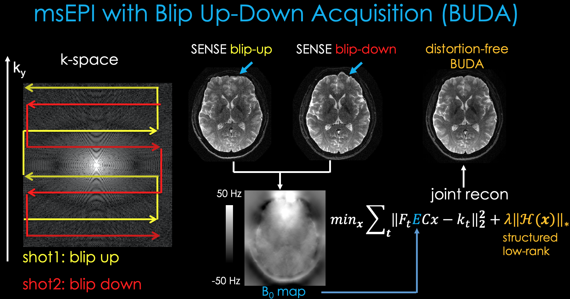
Blip-Up and -Down Acquisition (BUDA):
BUDA is a multi-shot EPI technique that acquires two shots of data with opposite phase encoding polarities [1,2]. Each individual shot is then reconstructed separately using parallel imaging, which are then processed with FSL's TOPUP [3] to estimate a field map. This field map is then incorporated into a joint parallel imaging reconstruction step where the two shots are reconstructed together, and their similarities are exploited using Hankel structured low-rank matrix constraints [4-6]. This obviates the need for navigator acquisitions since shot-to-shot phase differences can be directly estimated and accounted for. The k-space sampling patterns are staggered between the shots as shown below to provide complementary frequency coverage.
Generalized slice‐dithered enhanced resolution (gSlider):
gSlider is an RF encoding super-resolution technique for high, isotropic resolution diffusion imaging [7]. This makes 5 different acquisitions at e.g. 5 mm slice thickness and uses a different RF pulse in each of the acquisitions. With the knowledge of the RF slice profiles, it then becomes possible to solve an inverse problem to resolve a volume with 1 mm thick slices. Each RF-encoded thick slice image is reconstructed with parallel imaging, after which their background phase is estimated and removed using real-valued diffusion processing [8] for self-navigation. gSlider provides a significant SNR boost compared to standard EPI due to its volumetric encoding ability as seen below.

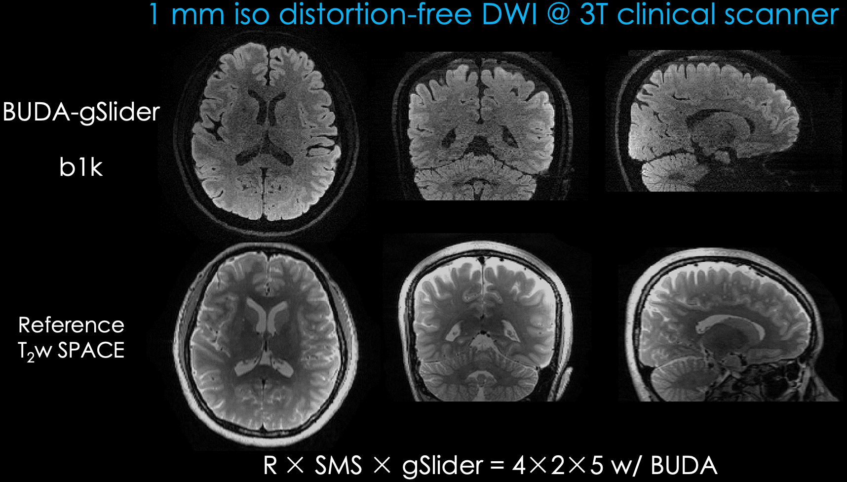
High-resolution, distortion-free diffusion imaging with BUDA-gSlider:
Combination of BUDA and gSlider strategies enable high-SNR, distortion-free diffusion imaging. This requires 10 shots of EPI per diffusion direction (2x AP/PA and 5x RF encoding). As shown below, this permits high geometric fidelity imaging on a clinical 3T system, and makes use of SMS acceleration with Blipped-CAIPI as well [9]. Further applications of BUDA-gSlider include high-resolution T2 and T2* mapping [10,11].
References:
- Liao, C., Bilgic, B., Tian, Q., Stockmann, J.P., Cao, X., Fan, Q., Iyer, S.S., Wang, F., Ngamsombat, C., Lo, W.C. and Manhard, M.K., 2021. Distortion‐free, high‐isotropic‐resolution diffusion MRI with gSlider BUDA‐EPI and multicoil dynamic B0 shimming. Magnetic resonance in medicine, 86(2), pp.791-803.
- Zahneisen, B., Aksoy, M., Maclaren, J., Wuerslin, C. and Bammer, R., 2017. Extended hybrid-space SENSE for EPI: Off-resonance and eddy current corrected joint interleaved blip-up/down reconstruction. NeuroImage, 153, pp.97-108.
- Andersson, J.L., Skare, S. and Ashburner, J., 2003. How to correct susceptibility distortions in spin-echo echo-planar images: application to diffusion tensor imaging. Neuroimage, 20(2), pp.870-888.
- Haldar, J.P., 2013. Low-rank modeling of local $ k $-space neighborhoods (LORAKS) for constrained MRI. IEEE transactions on medical imaging, 33(3), pp.668-681.
- Mani, M., Jacob, M., Kelley, D. and Magnotta, V., 2017. Multi‐shot sensitivity‐encoded diffusion data recovery using structured low‐rank matrix completion (MUSSELS). Magnetic resonance in medicine, 78(2), pp.494-507.
- Lee, D., Jin, K.H., Kim, E.Y., Park, S.H. and Ye, J.C., 2016. Acceleration of MR parameter mapping using annihilating filter‐based low rank hankel matrix (ALOHA). Magnetic resonance in medicine, 76(6), pp.1848-1864.
- Setsompop, K., Fan, Q., Stockmann, J., Bilgic, B., Huang, S., Cauley, S.F., Nummenmaa, A., Wang, F., Rathi, Y., Witzel, T. and Wald, L.L., 2018. High‐resolution in vivo diffusion imaging of the human brain with generalized slice dithered enhanced resolution: Simultaneous multislice (g S lider‐SMS). Magnetic resonance in medicine, 79(1), pp.141-151.
- Eichner, C., Cauley, S.F., Cohen-Adad, J., Möller, H.E., Turner, R., Setsompop, K. and Wald, L.L., 2015. Real diffusion-weighted MRI enabling true signal averaging and increased diffusion contrast. NeuroImage, 122, pp.373-384.
- Setsompop, K., Gagoski, B.A., Polimeni, J.R., Witzel, T., Wedeen, V.J. and Wald, L.L., 2012. Blipped‐controlled aliasing in parallel imaging for simultaneous multislice echo planar imaging with reduced g‐factor penalty. Magnetic resonance in medicine, 67(5), pp.1210-1224.
- Cao, X., Wang, K., Liao, C., Zhang, Z., Srinivasan Iyer, S., Chen, Z., Lo, W.C., Liu, H., He, H., Setsompop, K. and Zhong, J., 2021. Efficient T2 mapping with blip‐up/down EPI and gSlider‐SMS (T2‐BUDA‐gSlider). Magnetic Resonance in Medicine, 86(4), pp.2064-2075.
- Zhang, Z., Cho, J., Wang, L., Liao, C., Shin, H.G., Cao, X., Lee, J., Xu, J., Zhang, T., Ye, H. and Setsompop, K., 2022. Blip up‐down acquisition for spin‐and gradient‐echo imaging (BUDA‐SAGE) with self‐supervised denoising enables efficient T2, T2*, para‐and dia‐magnetic susceptibility mapping. Magnetic Resonance in Medicine.